Privately billed 3D Cone Beam CT, rebate can may be claimed with medicare by patient.
*Where Medicare is applicable
What is cone beam CT and how does it work?
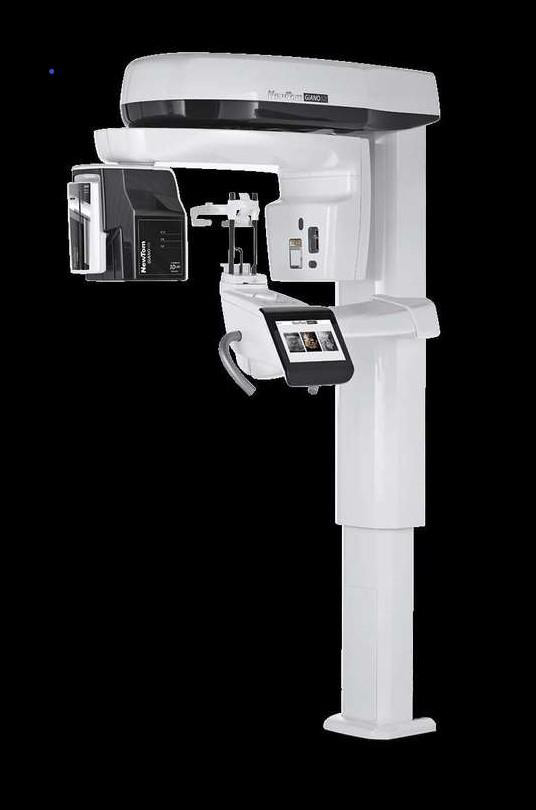
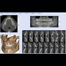
During a cone beam CT examination, the C-arm or gantry rotates around the head in a complete 360-degree rotation while capturing multiple images from different angles that are reconstructed to create a single 3-D image.
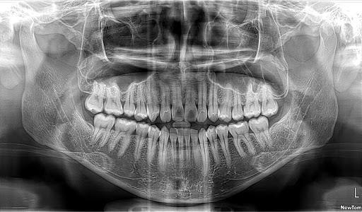
Cone Beam CT is a relatively new technique, which is mainly used to assess the jaws and teeth. Its main advantage compared to OPGs and other dental x-rays is that it provides three dimensional imaging (similar to conventional CT, but with a lower radiation dose). Kippax medical imaging uses the Cone Beam CT system, which provides high definition, three dimensional imaging to complement our other dental imaging services including
OPGs and conventional CT scanning. Cone Beam CT offers 3-D evaluation of dental anatomy and pathology: for example impacted teeth, pre-implant assessment, orthodontic assessment, and assessment for jaw and face surgery.
Medicare rebates apply for Medicare eligible patients if they are referred by their dental specialist or medical doctor. Medicare rebates do not apply to
referrals from general dentists.

Is cone beam CT Safe?
As outlined in the ALARA principle, every precaution should be taken to minimize radiation exposure. Radiation exposure from CBCT is up to 10 times less than that incurred from medical CT scanning, which exposes a patient to a dose of approximately 400 to 1000 µSv.
What is Cone beam useful for?

CBCT is increasingly being used by dental and maxillofacial surgeons to evaluate their patient’s anatomy with a 3D perspective. These indications include:
- Location of anatomical structures – mandibular canal, maxillary sinus,
- submandibular fossa
- Evaluation of the jaw for implant planning and for previously placed implants
- Evaluation of abnormalities in or affecting the bones – infections, cysts, tumours
- Evaluate extent of alveolar ridge resorption
- Impacted teeth
- Evaluation of bone structure and tooth orientation
- Alveolar fractures
- Assessment of the mandibular nerve prior to the removal of impacted teeth
- TMJ analysis
- Jaw lesions/tumours – relationships to other anatomical structures, expansion, erosions etc.
- Cephalometric analysis
- Oral surgery
- Endodontics/root canal
- Sleep apnea – Air way analysis
- Cleft palate
- Periodontal diseases
- Orthodontic planning
Your images and Report?
After your examination, you will be given a copy of the most pertinent images from your study. The three-dimensional image data will also be provided on a disc to the dentist or specialist who referred you, and can be used with other software, for example for implant planning.

A report will be given to you with the images, or sent directly back to your referring doctor by fax or email. KMI will store digital copies of all studies on our secure database for comparison with any future examinations.
Please bring any previous imaging with you for comparison.
It is important that you return to your doctor with your examination results. Whether they are normal or abnormal, your doctor needs to know promptly so that a management plan can be formulated
What should a Patient tell the radiographer before any radiological procedure ?
You will need to tell the radiographer if you are pregnant or suspect that you
might be pregnant
Cost Information
3D Cone Beam CT is privately billed, However, patients can submit their claim through medicare where applicable.
THE NEXT STEP
Please ensure that you have your Medicare card and referral with you and pop into our radiology clinic for your X-ray. If you have any questions at all please feel free to contact us, as we are here to help.

 Serving patients and Saving their lives